WORK & LIFTING INJURIES · MRI DECISION GUIDE · LOGANSPORT, IN
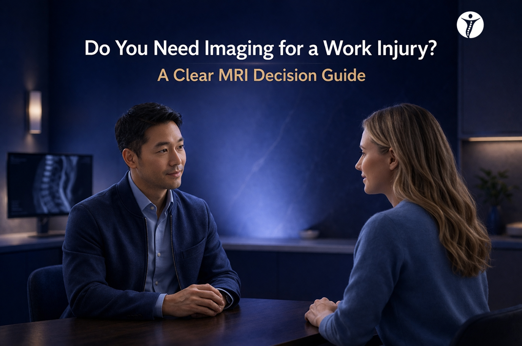
Do You Need Imaging for a Work Injury? A Clear MRI Decision Guide
Most work injuries don’t need an MRI right away—but some do. This guide shows you which is which.

Imaging can be valuable—but it’s not always the first best step. Most strains/sprains improve with the right plan and smart work modifications. If your injury is work-related, start with Work & Lifting Injuries. If symptoms involve your low back, see Low Back Pain Treatment.
- Clear “image now” red flags
- Timelines that actually match real recovery
- Simple next steps if you’re unsure
Educational only. Not medical advice. Seek urgent care for severe/worsening symptoms or red flags.
Quick Answer (The Simple Rule)
Most work injuries do not need an MRI right away. Imaging is most important when there are red flags, progressive neurologic symptoms, suspected fracture, or when the result will change decisions.

Image sooner if…
- There’s progressive weakness or worsening numbness
- You can’t bear weight or there’s a suspected fracture
- There are bowel/bladder changes or saddle numbness
- Severe trauma, rapidly worsening pain, or systemic symptoms
Conservative plan first if…
- No red flags
- Symptoms are stable or improving week-to-week
- Function is gradually returning (less guarding, better motion)
Red Flags (Image Now / Urgent Evaluation)
These aren’t common—but they matter. If any are present, err on the side of safety.
- Progressive weakness (foot drop, grip loss, can’t raise arm/leg like before)
- Loss of bowel/bladder control or saddle numbness
- Severe trauma (fall from height, major accident) or suspected fracture
- Fever with spinal pain, hot/red swollen joint, or feeling very unwell
- Rapidly worsening symptoms, or severe night pain that keeps escalating
If you’re unsure, start with Contact & Location and we’ll guide you to the safest next step.
Timeline: When Imaging Becomes More Useful
Imaging is most helpful when it changes the decision-making—not when it just adds labels.
0–2 weeks (early phase)
- Most strains/sprains are treated the same early: calm irritation + restore motion
- MRI often does not change the plan if there are no red flags
- Watch for: improving motion, fewer spikes, better sleep
2–6 weeks (rebuild phase)
- If you’re improving, keep progressing (this is where results compound)
- If you’re stuck (no progress) or worsening, consider re-evaluation and imaging discussion
6+ weeks (persistent limitation)
- Imaging is more likely to change decisions if function is still limited
- Especially if symptoms are nerve-y, strength is not returning, or pain is worsening
Progress markers that matter more than “pain today”
- You move more freely day-to-day
- Fewer “gotcha” spikes
- Sleep is improving
- Work tolerance is improving (even with modifications)
When Imaging Helps (and When It Often Doesn’t)
A simple table that keeps you out of “MRI just to see” traps.
| Imaging helps when… | Imaging often doesn’t help when… |
|---|---|
|
|
High-trust statement
We’re not anti-imaging—we’re pro-right-timing. The best time to image is when the result changes your next decision.
MRI vs X-ray vs CT (Simple)
Here’s the difference in plain language.
X-ray
Best for bones—fracture suspicion, major structural concerns, or certain joint issues.
MRI
Best for soft tissue—discs, nerves, ligaments. Most useful when symptoms are not improving, neurologic deficits are present, or results change decisions.
CT
Best for detailed bone imaging—sometimes used for complex fractures or when MRI isn’t possible.
MRI Words Explained (Don’t Panic)
Many MRI findings are common—even in people without pain. The key is whether they match your symptoms and exam.
Bulge vs herniation vs degeneration
These terms describe what the disc looks like—not how you’ll feel. A bulge can be painless; a small herniation can be painful; and degeneration is common with age. Read next: Disc Herniation vs. Bulge vs. Degeneration: What MRI Words Actually Mean.
“Tear” language
Imaging reports often use “tear” terms that sound scary. The real question is whether it matches your symptoms and function—and what your next best step is.
Best mindset
Imaging should be used to guide decisions—not to label you as “broken.” Your symptoms + exam drive the plan.
What to Do First (Without Guessing)
A simple action ladder that works for most non-red-flag work injuries.
Step 1: Reduce the spike (work modifications)
- Temporarily avoid the exact movement that triggered the flare
- Use shorter bouts and better positions rather than “powering through”
Step 2: Restore safe motion
- Gentle range of motion in pain-safe directions
- Stop repeatedly “testing” the painful movement
Step 3: Rebuild tolerance (the part that prevents re-injury)
- Light strength progression and capacity building
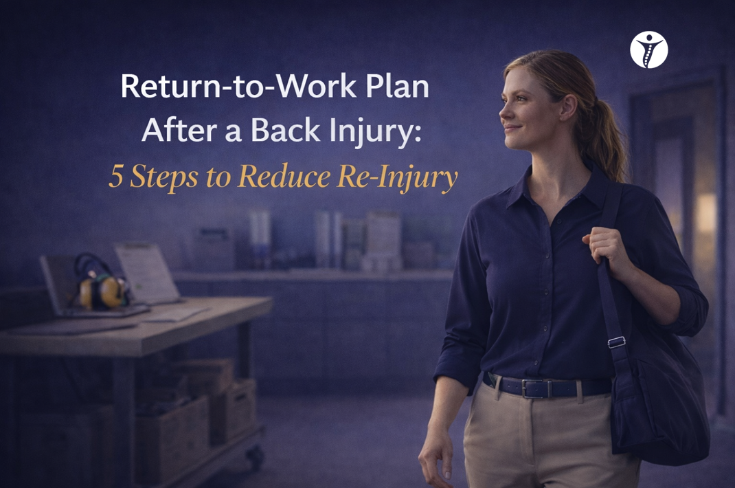
- Related: Return-to-Work Plan After a Back Injury: 5 Steps to Reduce Re-Injury
Step 4: Recheck milestones and decide on imaging if stalled
- If you’re not improving, we reassess the driver and next steps
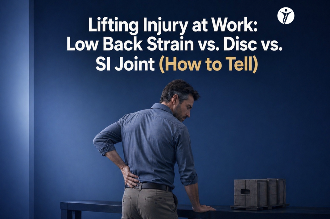
- Related: Lifting Injury at Work: Low Back Strain vs. Disc vs. SI Joint (How to Tell)
Work Injury Imaging FAQs
Quick answers—including “when to worry.”